The Process
Specimens
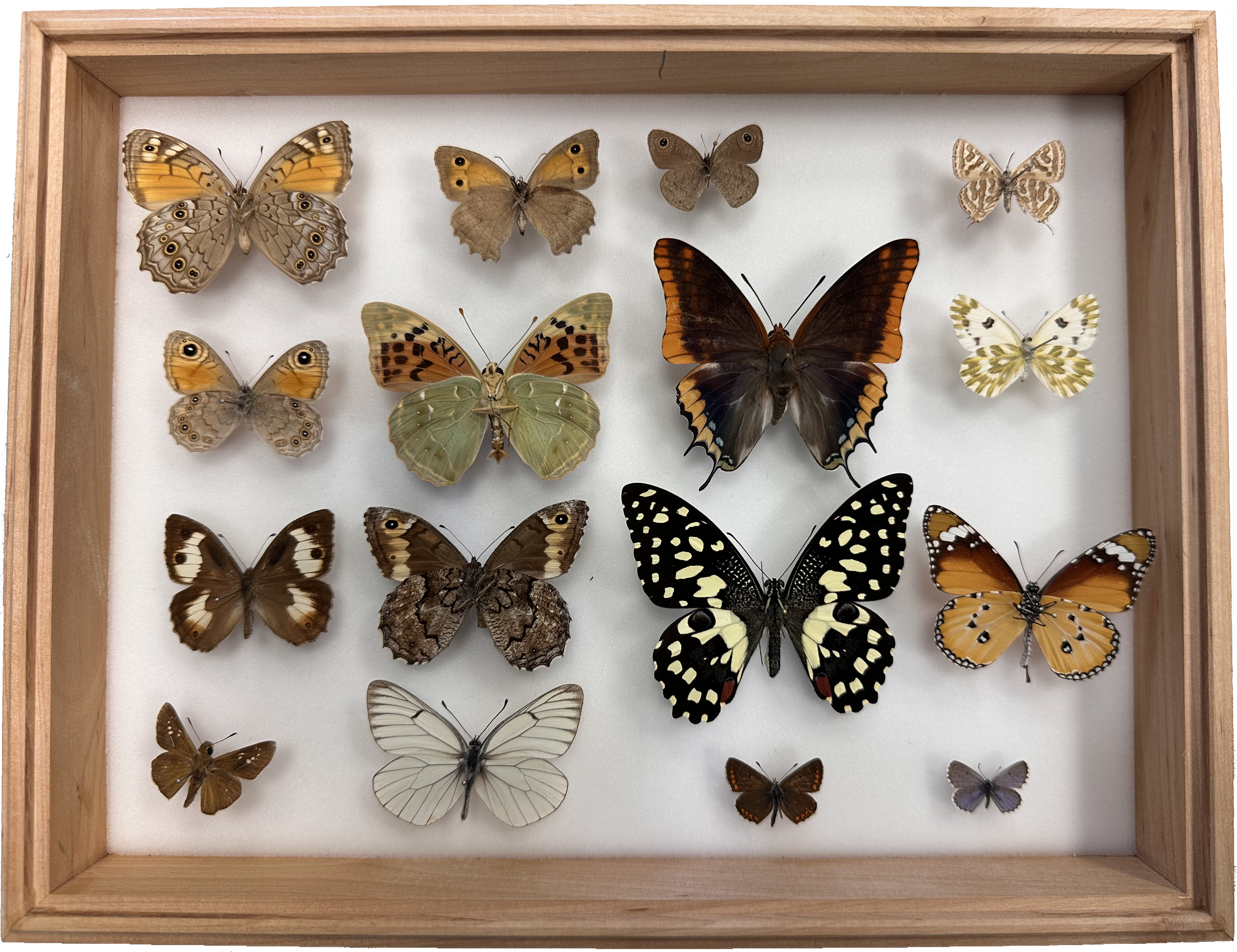
Butterfly specimens were obtained from curated private collections in Cyprus and from the Goulandris Natural History Museum in Greece. Together, they represent the full native butterfly fauna of Cyprus, along with selected Greek species. All specimens were already preserved, mounted on entomological pins, and stored in protective wooden collection boxes with transparent lids. Each was accompanied by coded labels containing species name, collection date, and location. Before scanning, specimens were positioned to ensure maximum visibility while minimizing the risk of damage.
3D Scanning Hardware
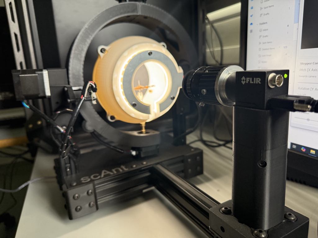
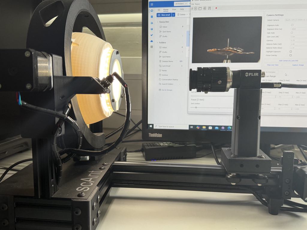
Digitization was carried out using the scAnt system, an open-source 3D scanner designed for small arthropods. The system combines a motorized camera rig with a three-axis rotation grid, controlled lighting, and automated software for image capture. Specimens were enclosed in a spherical chamber that ensured even illumination, supported by a custom LED flash system. Camera exposure, gain, and white balance were adjusted individually for each butterfly, with brighter specimens requiring shorter exposures and darker ones longer. This calibration helped preserve fine scale textures and subtle color patterns, especially in iridescent wings.
Scanning Workflow
Each scanning session began with initializing the scAnt system, connecting camera and motor controls, and setting up storage space. A single butterfly scan could exceed 200 GB, as images were captured in RAW format at high resolution (5472 × 3648 pixels). The process for each specimen involved:
- Defining movement ranges: Pitch, rotation, and focus step ranges were set to ensure full coverage of the specimen.
- Image capture: At each angle, stacks of images were taken with varying focus depths, producing a sharp, extended depth-of-field result.
- Image stacking: Multiple frames per viewpoint were merged into single composite images.
- Background removal: Automated algorithms cleaned each image by masking background elements, leaving only the specimen.
On average, setup and parameter adjustment took 10–15 minutes, while the full imaging process required about 3 to 3.5 hours per butterfly. The result was a complete set of stacked, cleaned images ready for 3D reconstruction.
3D Reconstruction using Photogrammetry
We generated high-resolution 3D models of the butterfly specimens using photogrammetry (Structure-from-Motion). This technique reconstructs geometry from overlapping photographs without physically touching the specimens, making it ideal for fragile material such as butterfly wings.
After scanning, we used 3DF Zephyr Lite to align the images, generate a sparse and dense point cloud, and build a triangulated mesh. The mesh was then cleaned in Blender to remove unwanted artifacts such as mounting pins and fill small gaps. The final step was texture mapping with the original photographs, adding realistic colors and details. Each reconstruction took about 60 minutes, plus 5–10 minutes of post-processing cleanup.
The resulting 3D models were exported in .OBJ and .STL formats for 3D printing and virtual reality applications. Cleaned models were then uploaded to Sketchfab where they can be explored interactively and embedded directly into this website.
This is the Sparse Point Cloud of the mesh, which is a 3D representation made up of a small number of points marking key features of the specimen. It provides a rough outline, forming the foundation for generating the dense point cloud and full 3D model.
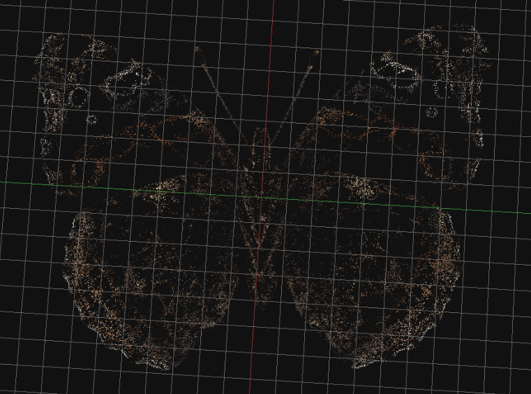
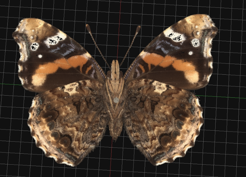
This is the Dense Point Cloud of the mesh, a detailed 3D representation with thousands of points covering the surface. It captures finer textures, shapes, and depth, serving as the basis for creating accurate meshes and textured models.
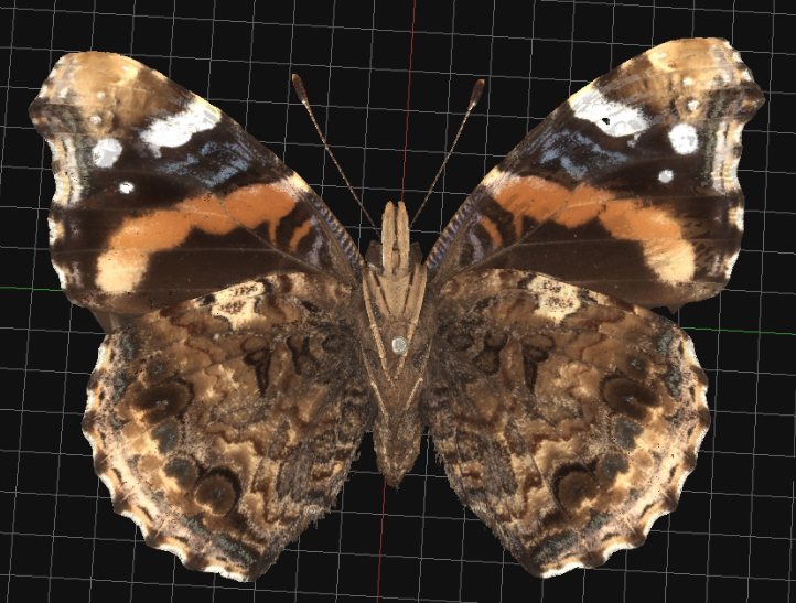
This is the final reconstruction of one of our specimens (Vanessa atalanta). It is the textured mesh built from the dense point cloud, providing a highly detailed 3D representation. This cleaned model is exported and uploaded to Sketchfab for interactive exploration.
Gaussian Splatting
In addition to photogrammetry, we created high-resolution 3D visualizations of the butterfly specimens using Gaussian Splatting. This cutting-edge method enables real-time 3D rendering by generating a continuous radiance field from overlapping photographs. Unlike traditional photogrammetry, which reconstructs polygonal meshes, Gaussian Splatting represents the scene as a collection of anisotropic 3D Gaussians. Each Gaussian encodes properties such as position, orientation, scale, color, and opacity, allowing for smooth, continuous, and highly photorealistic visualizations without the need for explicit surface triangulation.
We used the same scanning images and processed them with Polycam to align the photographs and prepare the input data for Gaussian Splatting. The resulting splats were further refined in Jawset PostShot to remove unwanted artifacts such as mounting pins and noise. The final visualizations preserve the appearance with realistic colors and details. Each reconstruction took about 40 minutes, plus 10–15 minutes of post-processing cleanup.
Gaussian splatting data (in .PLY format) can be provided upon request.
Metadata
Each digital specimen is paired with structured ecological metadata, stored alongside the images and models. Metadata includes taxonomy, distribution, wingspan, sexual dimorphism, life cycle and host plants, migration patterns, altitudinal range, and conservation status. By linking this information directly to the 3D models, the project combines accurate visual replicas with detailed biological context, creating a resource for research, education, and conservation.